Your brain is running right now at somewhere between 0.02 and 600 Hz, depending on what you’re doing. That’s not a metaphor. It’s a measurable electrical fact, and it’s the foundation of everything neuroscientists, clinicians, and sound therapists have built their work on.
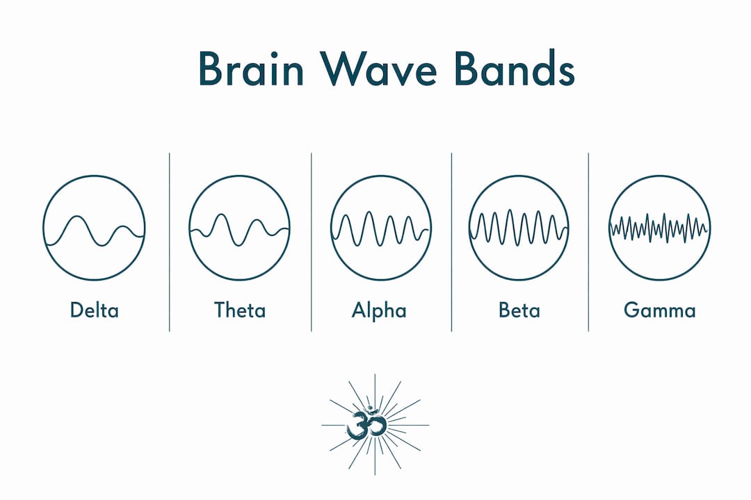
Brain waves are rhythmic electrical oscillations produced by synchronised neural activity; researchers primarily classify them by their frequency, measured in cycles per second (hertz). The standard scientific classification groups them into five core bands, with additional ranges extending the spectrum considerably further:
- Delta (δ) — deep sleep, tissue repair, unconscious processing
- Theta (θ) — drowsiness, creativity, early sleep stages
- Alpha (α) — relaxed wakefulness, the brain’s default background rhythm
- Sigma (σ) / sleep spindles — N2 sleep, memory consolidation
- Beta (β) — active thinking, focus, motor control
- Gamma (γ) — sensory integration, higher cognition
- High-frequency oscillations — very high frequency activity observed in advanced EEG research
- Infraslow oscillations — very slow brain activity dominant in preterm neonates, also present in non-REM sleep
The full electrophysiological spectrum of the brain spans approximately 0.02 to 600 Hz, though conventional clinical EEG typically focuses on the 0.5–70 Hz window.
What do different brain wave frequencies actually do?
Think of each frequency band less as a separate channel and more as a gear your brain shifts into depending on the task at hand. They overlap, interact, and coexist, which is precisely what makes them so fascinating to study.
Delta (0.5–4 Hz)
Delta waves are among the slowest brain waves and typically have a large amplitude. They predominate during deep, dreamless sleep and are prominent in frontocentral scalp regions. Physiologically, this period is associated with brain maintenance activities such as cellular repair and memory consolidation. When delta activity is observed during wakefulness, it can indicate neurological issues. Delta rhythms have also been implicated in sensorimotor functions related to stepping movements.
Theta (4–7 Hz)
Theta waves are commonly observed during drowsiness and early sleep stages, starting in frontocentral regions and moving backward as consciousness fades. Emotional arousal can increase frontal theta activity, especially in younger populations. Theta is often associated with a transitional state between wakefulness and sleep that fosters creative insight.
Alpha (8–12 Hz)
Alpha waves represent the brain’s resting rhythm, especially noted in the occipital region of adults. They become prominent in early childhood and typically remain stable across most of adult life. Closing the eyes often enhances alpha power. Slowing of alpha rhythms can indicate generalized cerebral dysfunction and is monitored clinically.

Sigma waves, or sleep spindles, are bursts of oscillatory activity observed during N2 sleep, reflecting thalamocortical circuit function. Slow and fast spindle variants have distinct roles; fast spindles relate to sensorimotor processing. Absence of typical spindle patterns in some children may indicate early developmental differences.
Beta waves are commonly observed during waking cognition, predominating in frontal and central brain regions. They are associated with active thinking, problem solving, focused attention, and motor control, including synchronization between motor cortex and spinal activity during sustained muscle use.
Gamma (30–80 Hz)
Gamma waves were first recorded in the visual cortex of monkeys and are now known to occur across the premotor, parietal, temporal, and frontal cortices. They’re associated with sensory processing and cognition, and the cortico-basal-ganglia-thalamo-cortical loop communicates using this rhythm. Elevated beta-gamma phase-amplitude coupling over the sensorimotor cortex is consistently observed in Parkinson’s disease, reflecting pathological neural entrainment in the basal ganglia.
High-frequency oscillations, including ripples and fast ripples, represent advanced EEG features under research. These patterns show promise as biomarkers for epilepsy surgical outcomes, with increasing clinical relevance due to advancements in digital signal processing enabling their detection.
Pro Tip: Brain wave bands are not rigid containers. As Britannica’s neural oscillation reference notes, dominant activity guides interpretation rather than absolute frequency cutoffs. Multiple oscillations coexist and interact dynamically at any given moment.
How do scientists actually measure brain wave frequencies?
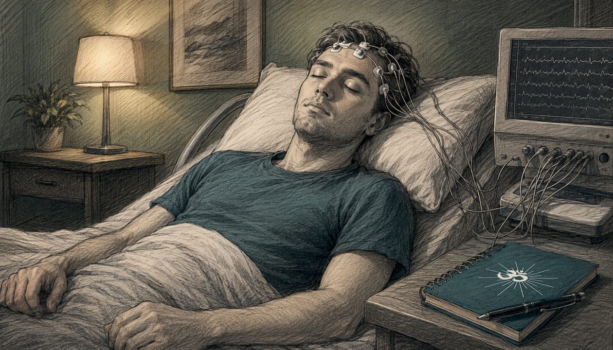
The primary tool is electroencephalography (EEG), a technique that places electrodes on the scalp to detect the summed electrical activity of millions of synchronised neurons beneath. A single neuron’s electrical signal is far too small to register outside the skull. What EEG captures is the constructive interference of thousands of neurons firing in coordinated patterns, producing measurable oscillations.
The standard clinical EEG pipeline works roughly like this:
- Signal acquisition: Electrodes (typically 19–256 channels) record raw electrical potential from the scalp surface.
- Bandpass filtering: Filters isolate specific frequency ranges, removing artefacts from muscle movement, eye blinks, and electrical interference.
- Spectral analysis: Techniques such as Fast Fourier Transform (FFT) convert the time-domain signal into a frequency-domain representation, showing the power at each Hz.
- Band power quantification: Researchers calculate the relative or absolute power within each frequency band to characterise brain states.
- Clinical or research interpretation: Patterns are compared against normative databases, adjusted for age, electrode location, and task condition.
Conventional clinical EEG focuses on 0.5 to 70 Hz using bandpass filtering. Digital signal processing advances have pushed this considerably further, revealing infraslow oscillations below 0.5 Hz and high-frequency oscillations up to 600 Hz. Simultaneous recording of direct current shifts, infraslow oscillations, local field potentials, and high-frequency activity from the same site is now being explored for preclinical epilepsy research, potentially offering biomarkers for precise seizure onset zone delineation.
Sound therapy, brain oscillations, and the work of Robert Emery and Moritz Schneider
Brain waves don’t operate in isolation from the rest of the body. Research into the frequency architecture of brain and body oscillations suggests that brain oscillations and body rhythms, including heart rate and breathing, form a coupled hierarchical system. The centre frequency of the traditional EEG bands follows a binary doubling relationship: delta at roughly 2.5 Hz, theta at 5 Hz, alpha at 10 Hz, beta at 20 Hz, and gamma at 40 Hz. Average resting heart rate fits neatly into this hierarchy as a foundational oscillator. Breathing frequency clusters at distinct peaks around 0.07, 0.15, and 0.30 Hz, all within predicted bands of the same system.
This is the scientific territory that composers Robert Emery and Moritz Schneider work within. Both bring serious musical and technical credentials to Orchestralmeditations’ recordings. Emery, a composer and producer with deep experience in orchestral and cinematic music, and Schneider, whose background spans classical composition and electronic sound design, collaborate on tracks that layer binaural beats, theta frequencies, and Solfeggio-based tones within full orchestral arrangements recorded at Abbey Road Studios with the National Philharmonic. The intent is not decorative. It’s to create soundscapes that influence brain wave states by aligning auditory input with the body’s own oscillatory rhythms.
The phase-amplitude coupling of slower and faster brain waves is a key principle here. When a slower frequency modulates the envelope of a faster one, it creates conditions for cross-frequency communication within the brain. Emery and Schneider’s compositions exploit this by embedding frequency layers that correspond to theta and alpha ranges within broader orchestral textures, supporting the transition from active beta-dominant wakefulness into deeper meditative states.
Key applications of brain wave frequency science in sound therapy and beyond:
- Meditation and relaxation: Alpha and theta entrainment through binaural beats and tuned soundscapes supports the shift from beta-dominant wakefulness into calmer states.
- Sleep support: Delta-range audio cues may assist the transition into deep sleep stages, relevant for insomnia research.
- Cognitive enhancement: Gamma-frequency stimulation is being investigated for its role in sensory integration and attention.
- Clinical neurology: Altered frequency patterns are observed in ADHD, Parkinson’s disease, epilepsy, and dementia, making EEG frequency analysis a diagnostic tool.
- Neurofeedback: Real-time EEG feedback trains individuals to modulate their own frequency patterns, with applications in anxiety, ADHD, and peak performance.
- Music therapy: Research into music therapy for dementia highlights how rhythmic auditory stimulation interacts with neural oscillations to support cognitive and emotional function.
Orchestralmeditations’ library, produced by Emery and Schneider, sits at the intersection of these applications. The recordings are available through the meditation music library, where frequency-layered orchestral compositions are designed for both casual listeners and those with a more specific therapeutic or meditative goal.

How brain wave classification got its Greek letters
The naming of brain waves is one of those delightful quirks of scientific history where convention stuck simply because it worked. Hans Berger, the German psychiatrist who recorded the first human EEG in 1924, named the dominant rhythm he observed “alpha” after the first letter of the Greek alphabet. When he identified a faster rhythm during mental activity, he called it “beta.” The convention of using Greek letters for subsequent bands followed naturally, though not always in alphabetical order of discovery.
Delta was named by W. Grey Walter in the 1930s, who used it to describe the slow waves he associated with sleep and brain lesions. Theta followed, and gamma came considerably later as high-frequency recording technology improved. Sigma, used for sleep spindles, reflects the waveform’s characteristic spindle shape rather than its position in the Greek alphabet.
The standardised classification used in EEG research today, with frequency as the primary organising principle, was formalised through decades of clinical neurophysiology. The International Federation of Clinical Neurophysiology (IFCN) has played a central role in establishing and maintaining these conventions, ensuring that an alpha rhythm recorded in London means the same thing as one recorded in Tokyo. Frequency remains the most commonly used method to classify EEG waveforms, with Greek letter naming tied directly to Hz ranges rather than to any functional or anatomical criterion.
One subtlety worth noting: the boundaries between bands are not universally agreed upon. Some sources place the lower limit of alpha at 8 Hz, others at 7.5 Hz. Beta’s upper boundary varies between 30 and 35 Hz depending on the research tradition. The neural oscillation literature treats these as flexible, overlapping ranges rather than hard cutoffs, which is scientifically more accurate and practically more useful for anyone designing neurofeedback protocols or sound therapy frequencies.
Key takeaways
Brain wave frequency bands, measured in hertz via EEG, each correspond to distinct cognitive and physiological states, spanning a full electrophysiological spectrum from approximately 0.02 to 600 Hz.
| Point | Details |
|---|---|
| Five core frequency bands | Delta (0.5–4 Hz), Theta (4–7 Hz), Alpha (8–12 Hz), Beta (13–30 Hz), and Gamma (30–80 Hz) form the standard EEG classification. |
| Extended spectrum matters | Infraslow oscillations (<0.5 Hz) and high-frequency oscillations (>80 Hz) carry physiological and pathological significance beyond the clinical EEG window. |
| Alpha is the brain’s baseline | The posterior alpha rhythm (8–12 Hz) defines normal adult EEG and remains stable from age three into the ninth decade of life. |
| Bands overlap dynamically | Frequency boundaries are flexible; dominant activity guides interpretation rather than absolute Hz cutoffs. |
| Sound therapy applies this science | Composers Robert Emery and Moritz Schneider layer binaural beats and theta frequencies in orchestral recordings to align auditory input with brain and body oscillatory rhythms. |
FAQ
What Hz are brain waves?
Brain waves span approximately 0.02 to 600 Hz across the full electrophysiological spectrum. The five clinically recognised bands are Delta (0.5–4 Hz), Theta (4–7 Hz), Alpha (8–12 Hz), Beta (13–30 Hz), and Gamma (30–80 Hz), with additional infraslow and high-frequency oscillations beyond these ranges.
What does 432 Hz do to your brain?
432 Hz is an audio pitch, not a brain wave frequency. Brain waves are measured in cycles per second of electrical oscillation within a range up to about 600 Hz; 432 Hz refers to the tuning of a musical note. No peer-reviewed EEG research establishes a direct causal link between listening to music tuned to 432 Hz and a specific measurable change in brain wave frequency bands.
What does 10,000 Hz do to your brain?
10,000 Hz is an audio frequency in the upper range of human hearing, well above any brain wave frequency band. Brain waves top out at around 600 Hz in research contexts; 10,000 Hz as a sound stimulus may influence auditory cortex processing, but it does not correspond to any recognised brain wave frequency classification.
What brain wave frequencies are associated with high cognitive performance?
Gamma oscillations (30–80 Hz) are most consistently linked to sensory integration, attention, and higher cognitive processing. Beta activity (13–30 Hz) supports active focus and working memory. Research into gamma-frequency stimulation is ongoing, with particular interest in its role in conditions such as Alzheimer’s disease and attention disorders.